This is default featured post 1 title

Go to Blogger edit html and find these sentences.Now replace these sentences with your own descriptions.This theme is Bloggerized by Lasantha Bandara - Premiumbloggertemplates.com.

This is default featured post 2 title

Go to Blogger edit html and find these sentences.Now replace these sentences with your own descriptions.This theme is Bloggerized by Lasantha Bandara - Premiumbloggertemplates.com.

This is default featured post 3 title

Go to Blogger edit html and find these sentences.Now replace these sentences with your own descriptions.This theme is Bloggerized by Lasantha Bandara - Premiumbloggertemplates.com.

This is default featured post 4 title

Go to Blogger edit html and find these sentences.Now replace these sentences with your own descriptions.This theme is Bloggerized by Lasantha Bandara - Premiumbloggertemplates.com.

This is default featured post 5 title

Go to Blogger edit html and find these sentences.Now replace these sentences with your own descriptions.This theme is Bloggerized by Lasantha Bandara - Premiumbloggertemplates.com.

Tampilkan postingan dengan label Double-Stranded DNA Viruses (FamilyF Who Have Not Marked). Tampilkan semua postingan

Senin, 17 Oktober 2011

Virus Famili Tectiviridae

The Tectiviridae is a family of double stranded DNA viruses that infect bacteria and archea. Tectiviridae have no head-tail structure, but are capable of producing tail-like tubes of ~ 60 x 10 nm upon adsorption or after chloroform treatment. The name is derived from Latin tectus (meaning 'covered').

There is a single genus in this family - genus Tectivirus.

The type species is Enterobacteria phage PRD1.
Virology

This family of viruses consist of virons with double layers capsids that have apical spikes extending ~20 nanometers (nm) and an unusual internal lipid envelope around the nucleoprotein.

The capsid is nonenveloped and has a diameter of 63 nm icosahedron structure. The capsid shells are composed of two layers - an inner and outer capsid.

The inner capsid shell consist of a 5-6 nm flexible shell made from a lipoprotein vesicle whereas the outer capsid is made up of a smooth, rigid 3 nm thin protein shell. The outer shell has a pseudo T = 25 symmetry and consists of 240 capsid proteins trimers.

The genome is a single molecule of linear double stranded DNA of 15 kilobases in length. It forms a tightly packed coil and encodes several structural proteins. It encodes ~30 proteins that are transcribed in operons.

At least 9 structural proteins are present in the viron.

The genome is ~66 megaDaltons in weigh and constitutes 14-15% of the virion by weight. Lipids constitute a further 15% by weight. CarbohydratLife cycle

After adsorbion to the host's cells surface the viron extruds a tail-tube structure through a vertex for genome delivery into the host.

Capsid proteins polymerize around a lipoprotein vesicle translocated in the cytoplasm by virion assembly factors.

Mature virons are released by lysis.es are not present.

Virus Famili Rudiviridae

The Rudivirus (members of the family Rudiviridae) are unenveloped, stiff-rod-shaped viruses with linear dsDNA genomes, that infect hyperthermophilic archaea of the kingdom Crenarchaeota. The study of crenarchaeal viruses is still incipient. Our knowledge of their biology and basic molecular processes, including infection, virus-host interactions, DNA replication and packaging, as well as transcription regulation, is somewhat limited.

Rudivirus are promising candidates to become a general model for detailed studies of archaeal virus biology. These are indeed easily maintained under laboratory conditions and can be obtained in sufficient yields, unlike many other archaeal viruses.

The family name derives from the Latin rudis, thin rod, referring to the virion shape.
Taxonomy

    Main species
        Sulfolobus islandicus rod-shaped virus 1, SIRV1; genome sequence accession no. AJ414696.
        Sulfolobus islandicus rod-shaped virus 2, SIRV2; genome sequence accession no. AJ344259.
    Other species
        Acidianus rod-shaped virus 1, ARV1; genome sequence accession no. AJ875026.
        Stygiolobus rod-shaped virus, SRV, genome sequence accession no. FM164764.

The two main species, viruses SIRV1 and SIRV2, were produced by colony-cloned Sulfolobus islandicus strains. The two strains were isolated from samples taken in 1994 from different solfataric fields in Iceland, the Kverkfjöll and Hveragerdi which are separated by a distance of 250 km. These Icelandic solfataric acidic hot springs reach a temperature of 88°C and pH 2.5. As for its stability in many hosts, SIRV2 is a better candidate for the type species than SIRV1.

Acidianus rod-shaped virus 1, ARV1, the first member of the family Rudiviridae infecting hyperthermophilic archaea of the genus Acidianus, was isolated from a hot spring in Pozzuoli, Italy in 2005.

The Stygiolobus rod-shaped virus, SRV, which infects a hyperthermophilic Stygiolobus species, was isolated from a hot spring in the Azores, Portugal in 2008

Structure
Virions are non-enveloped, consisting of a tube-like superhelix formed by dsDNA and the major structural protein, with plugs at each end to which three tail fibers are anchored. These tail fibers appear to be involved in adsorption onto the host cell surface and are formed by one of the minor structural proteins.

Both Sulfolobus islandicus rod-shaped viruses are stiff rods of about 23 nm in width, but differing in length — SIRV1 is about 830 nm and SIRV2 is about 900 nm long. They present a central channel of approx. 6 nm that encapsidates the DNA genome. At each terminus of the rod there is a plug of approx. 48 nm in length and 6 nm in diameter that fills the terminal portion of the cavity, together with three tail fibres of approx. 28 nm in length.

Acidianus rod-shaped virus 1 is 610 nm long and 22 nm wide, also has the three tail fibers protruding at each end and the same central channel encapsidating the genome.

The Stygiolobus rod-shaped virus displays a similar rod-shaped morphology, sizing 702 nm by 22 nm.
Genome

The rudiviral genome is composed of linear dsDNA and ranges from of 24 kb (ARV1) to 35 kb (SIRV2).The two strands of the linear genomes are covalently linked and, at both ends of the genome, there are inverted terminal repeats. The Sulfolobus rudiviruses size up to 32.3 kbp for SIRV1 and 35.8 kbp for SIRV2, with inverted terminal repeats of 2029 bp at the ends of the linear genome. The G+C content of both genomes is extremely low, of only 25%, whereas the genome of Sulfolobus solfataricus (the sequenced genome closest to the virus host) hits 37%.

The genome sequence and composition of ARV1 differs strongly from those of the Sulfolobus rudiviruses. ARV1 has a genome of 24,655 bp, including 1365 bp inverted terminal repeats at both ends.

SRV shows sufficient genomical differences from the other rudiviruses to warrant its classification as a novel species. Its genome totals 28,096 bp and presents inverted terminal repeats of 1,030 bp.

Although the sequences of the inverted terminal repeats of the rudiviruses are different, they all carry the motif AATTTAGGAATTTAGGAATTT near the genome ends which may constitute a signal for the Holliday junction resolvase and DNA replication.

Transcriptional Patterns and Transcription Regulation
The transcriptional patterns of the rudiviruses SIRV1 and SIRV2 are relatively simple, with few temporal expression differences. Contrastingly, at least 10% of its genes were predicted to have of different DNA binding motifs in the proteins they code and were assigned to be putative transcriptional regulators. A high proportion of viral genes coding for DNA binding proteins with the ribbon-helix-helix (RHH) DNA binding motifs has been suggested. The abundance of genes coding for proteins belonging to the RHH superfamily present in the genomes of crenarchaea and their viruses could underline the important role of these proteins in host and viral gene transcription regulation under harsh conditions.

Protein SvtR was the first crenarchaeal RHH regulator characterized in details and also the first viral coded transcriptional regulators within the Archaeal domain. It strongly represses the transcription of the minor structural protein and, to a lesser extent, of its own gene. The structure is very similar to that of bacterial RHH proteins despite the low sequence similarity, such as CopG, a bacterial plasmid copy number control regulator.

A Sulfolobus islandicus coded transcription activator, Sta1, as also been shown to activate transcription of several viral genes

Sulfolobus islandicus rod-shaped virus 2 (SIRV2) is a lytic virus that kills the host cell as a consequence of elaborated mechanisms orchestrated by the virus. Massive degradation of the host chromosomes occurs because of virus infection and virion assembly occurs in the cytoplasm. Virions are released from the host cell through a mechanism that involves the formation of specific cellular structures.

Potential applications in Nanotechnology
SIRV2 can act as a template for site-selective and spatially controlled chemical modification. Both the ends and the body of the virus, or the ends only, can be chemically addressed, thus SIRV2 can be regarded as a structurally unique nanobuilding block.

Virus Famili Poxviridae

Poxviruses (members of the family Poxviridae) are viruses that can, as a family, infect both vertebrate and invertebrate animals.

Four genera of poxviruses may infect humans: orthopox, parapox, yatapox, molluscipox. Orthopox: smallpox virus (variola), vaccinia virus, cowpox virus, monkeypox virus; Parapox: orf virus, pseudocowpox, bovine papular stomatitis virus; Yatapox: tanapox virus, yaba monkey tumor virus; Molluscipox: molluscum contagiosum virus (MCV). The most common are vaccinia (seen on Indian subcontinent) and molluscum contagiousum, but monkeypox infections are rising (seen in west and central African rainforest countries).
Structure

Poxviridae viral particles (virions) are generally enveloped (external enveloped virion- EEV), though the intracellular mature virion (IMV) form of the virus, which contains different envelope, is also infectious. They vary in their shape depending upon the species but are generally shaped like a brick or as an oval form similar to a rounded brick because they are wrapped by the endoplasmic reticulum. The virion is exceptionally large, its size is around 200 nm in diameter and 300 nm in length and carries its genome in a single, linear, double-stranded segment of DNA. By comparison, Rhinovirus is 1/10 as large as a typical Poxviridae virion.

Replication
Replication of the poxvirus involves several stages. The first thing the virus does is to bind to a receptor on the host cell surface; the receptors for the poxvirus are thought to be Glycosaminoglycans (GAGs). After binding to the receptor, the virus enters the cell where it uncoats. Uncoating of the virus is a two step process. Firstly the outer membrane is removed as the particle enters the cell; secondly the virus particle (without the outer membrane) is uncoated further to release the core into the cytoplasm. The pox viral genes are expressed in two phases. The early genes are expressed first. These genes encode the non-structural protein, including proteins necessary for replication of the viral genome, and are expressed before the genome is replicated. The late genes are expressed after the genome has been replicated and encode the structural proteins to make the virus particle. The assembly of the virus particle occurs in the cytoskeleton of the cell and is a complex process that is poorly understood but is currently being researched. Considering the fact that this virus is large and complex, replication is relatively quick taking approximately 12 hours until the host cell dies by the release of viruses.

The replication of poxvirus is unusual for a virus with double-stranded DNA genome (dsDNA) because it occurs in the cytoplasm[4] Poxvirus encodes its own machinery for genome transcription, a DNA dependent RNA polymerase, which makes replication in the cytoplasm possible. Most dsDNA viruses require the host cell's proteins to perform transcription. These host proteins are found in the nucleus, and therefore most dsDNA viruses carry out a part of their infection cycle within the host cell's nucleus.

axonomy
The name of the family, Poxviridae, is a legacy of the original grouping of viruses associated with diseases that produced poxes in the skin. Modern viral classification is based on phenotypic characteristics; morphology, nucleic acid type, mode of replication, host organisms, and the type of disease they cause. The smallpox virus remains as the most notable member of the family.

The following genera are currently included here:

    Subfamily Chordopoxvirinae
        Genus Avipoxvirus; type species: Fowlpox virus; species: Canarypox virus, Fowlpox virus, Juncopox virus, Mynahpox virus, Pigeonpox virus, Psittacinepox virus, Quailpox virus, Sparrowpox virus, Starlingpox virus, Turkeypox virus
        Genus Capripoxvirus; type species: Sheeppox virus; species: Goatpox virus, Lumpy skin disease virus, Sheeppox virus
        Genus Cervidpoxvirus; type species: Deerpox virus W-848-83; species: Deerpox virus W-848-83
        Genus Leporipoxvirus; type species: Myxoma virus; species: Hare fibroma virus, Myxoma virus, Rabbit fibroma virus, Squirrel fibroma virus
        Genus Molluscipoxvirus; type species: Molluscum contagiosum virus; species: Molluscum contagiosum virus
        Genus Orthopoxvirus; type species: Vaccinia virus; species: Camelpox virus, Cowpox virus, Ectromelia virus, Monkeypox virus, Raccoonpox virus, Taterapox virus, Vaccinia virus, Variola virus, Volepox virus
        Genus Parapoxvirus; type species: Orf virus; species: Bovine papular stomatitis virus, Orf virus, Parapoxvirus of red deer in New Zealand, Pseudocowpox virus
        Genus Suipoxvirus; type species: Swinepox virus; species: Swinepox virus
        Genus Unassigned; species: Squirrel poxvirus
        Genus Yatapoxvirus; type species: Yaba monkey tumor virus; species: Tanapox virus, Yaba monkey tumor virus
    Subfamily Entomopoxvirinae
        Genus Alphaentomopoxvirus; type species: Melolontha melolontha entomopoxvirus; species: Anomala cuprea entomopoxvirus, Aphodius tasmaniae entomopoxvirus, Demodema boranensis entomopoxvirus, Dermolepida albohirtum entomopoxvirus, Figulus subleavis entomopoxvirus, Geotrupes sylvaticus entomopoxvirus, Melolontha melolontha entomopoxvirus
        Genus Betaentomopoxvirus; type species: Amsacta moorei entomopoxvirus 'L'; species: Acrobasis zelleri entomopoxvirus 'L', Amsacta moorei entomopoxvirus 'L', Arphia conspersa entomopoxvirus 'O', Choristoneura biennis entomopoxvirus 'L', Choristoneura conflicta entomopoxvirus 'L', Choristoneura diversuma entomopoxvirus 'L', Choristoneura fumiferana entomopoxvirus 'L', Chorizagrotis auxiliars entomopoxvirus 'L', Heliothis armigera entomopoxvirus 'L', Locusta migratoria entomopoxvirus 'O', Oedaleus senigalensis entomopoxvirus 'O', Operophtera brumata entomopoxvirus 'L', Schistocera gregaria entomopoxvirus 'O'
        Genus Gammaentomopoxvirus; type species: Chironomus luridus entomopoxvirus; species: Aedes aegypti entomopoxvirus, Camptochironomus tentans entomopoxvirus, Chironomus attenuatus entomopoxvirus, Chironomus luridus entomopoxvirus, Chironomus plumosus entomopoxvirus, Goeldichironomus haloprasimus entomopoxvirus
        Genus Unassigned; species: Diachasmimorpha entomopoxvirus

The prototype of poxvirus family is vaccinia virus, which has been used as a successful vaccine to eradicate smallpox virus. Vaccinia virus is also used as an effective tool for foreign protein expression to elicit strong host immune response. Vaccinia virus enters cells mainly by cell fusion, although currently the receptor is not known. Virus contains three classes of genes, early, intermediate and late, that are transcribed by viral RNA polymerase and associated transcription factors. Vaccinia virus replicates its genome in cytoplasm of the infected cells and after late gene expression virion morphogenesis produces IMV that contains envelope, although the origin of the envelope membrane is still unknown. IMV is transported to Golgi to be wrapped additional two membrane to become intracellular enveloped virus (IEV). IEV transports along microtubules to reach cell periphery and fuse with plasma membrane to become cell-associated enveloped virus (CEV) that triggers actin tails on cell surfaces or is releared as EEV.
Diseases caused by pox viruses, especially smallpox, have been known about for centuries. One of the earliest suspected cases is that of Egyptian pharaoh Ramses V who is thought to have died from smallpox nearly 2000 years BCE.

Smallpox was thought to have been transferred to Europe around the early 8th century and then to the Americas in the early 16th century. It is widely accepted that the main defeat of the Aztecs was due to a smallpox epidemic and within two years over 3.2 million Aztecs died. This death toll can be attributed to the American population's complete lack of sensitization to the virus as children. A century after Edward Jenner showed that the less potent cow pox could be used to effectively vaccinate against the more deadly smallpox, a worldwide effort to vaccinate everyone against smallpox began with the ultimate goal to rid the world of the plague-like epidemic. The World Health Organization (WHO) declared the virus officially eradicated in 1977 and after nine years, in 1986, all virus samples were destroyed or transferred to two approved WHO reference labs: at the headquarters of the federal Centers for Disease Control and Prevention (the C.D.C.) in Atlanta, Georgia (the United States) and at the Institute of Virus Preparations in Moscow.  Post September 11, 2001 the American and UK governments have had increased concern over the use of smallpox, or a small pox like disease, in bio-terrorism.

Virus Famili Polyomaviridae

Polyomaviridae es una familia de virus que contiene un único género, Polyomavirus, que infecta animales. Los poliomavirus tienen un genoma ADN de cadena doble y por lo tanto se incluyen en el Grupo I de la Clasificación de Baltimore. El genoma es circular, de 5000 pares de bases. Los virus tienen un tamaño pequeño, 40-50 nm de diámetro, forma icosaédrica y carecen de envoltura de lipoproteína. Son potencialmente oncogénicos (causantes de tumores); frecuentemente persisten como infecciones latentes en un hospedante sin causar enfermedad, pero pueden producir tumores en un huésped de especie diferente, o en un huésped con un ineficiente sistema inmunitario. El nombre polioma refiere a la habilidad del virus de producir múltiples (poli) tumores (oma).

Se conocen cinco especies de poliomavirus que infectan humanos:

    Poliomavirus BK, que puede infectar el sistema respiratorio, riñones y cerebro, puede producir cistitis hemorrágica o una nefropatía
    Poliomavirus JC, a veces mortal al causar leucoencefalopatía multifocal progresiva.

Ambos virus están  muy extendidos: el 80 por ciento de la población adulta

en los Estados Unidos tienen anticuerpos frente a BK y JC.

    Dos virus recientemente descubiertos, Poliomavirus KI (Instituto Karolinska)1 y Poliomavirus WU (Universidad de Washington)2 están estrechamente relacionados entre sí y han sido aislados de las secreciones respiratorias.
    En enero de 2008 se describió una nueva especie, Poliomavirus de células de Merkel como el probable agente causal de cáncer de piel de Merkel.3

El Virus vacuolante del simio 40 se replica en los riñones de monos sin causar enfermedad, pero causa sarcomas en hámsters. Se desconoce si puede causar enfermedades en los seres humanos, lo que ha causado preocupaciones, ya que el virus puede haber sido introducido en la población general en la década de 1950 a través de una vacuna contra la polio contaminada. Una hipótesis similar postula que esta vacuna pudo haber sido la causa de la transmisión del virus del sida de los chimpancés a los humanos. El Virus de la enfermedad del polluelo del periquito es una causa frecuente de muerte entre las aves enjauladas.

El género Poliomavirus solía ser uno de los dos géneros dentro de la familia ahora obsoleta Papovaviridae (el otro género, el virus del papiloma, ahora se clasifica en su propia familia, Papillomaviridae).
Replicación

Antes de la replicación del genoma tienen lugar los procesos de fijación, entrada y liberación de la cubierta. Actualmente se desconocen los receptores celulares para los poliomavirus, sin embargo, la fijación del poliomavirus a la célula huésped es mediada por la proteína viral 1 (VP1). Esto se ha demostrado al comprobarse que los anticuerpos anti-VP1 previenen la unión del poliomavirus a la célula huésped.4

A continuación los viriones son endocitados y posteriormente transportados directamente al núcleo en vacuolas endocíticas en donde se produce la liberación de la cubierta del virus.

Los poliomavirus se replican en el núcleo de la célula huésped. Son capaces de utilizar la maquinaria genómica del huésped puesto que su estructura genómica es homóloga a la del huésped mamífero. La replicación viral se produce en dos fases distintas: expresión de genes inicial y final, separadas por la replicación del genoma.

La expresión de genes inicial es la responsable de la síntesis de proteínas no estructurales. Puesto que los poliomavirus confían en el huésped para el control de la expresión génica, la función de las proteínas no estructurales es la de regular los mecanismos celulares. Cerca del terminal N del genoma del poliomavirus hay elementos potenciadores que inducen la activación y transcripción de una molécula conocida como el antígeno T. El ARNm inicial codifica antígenos T que son producidos por la ARN polimerasa II del huésped. El antígeno T autorregula el ARNm inicial, que posteriormente conduce a niveles elevados del antígeno T. A altas concentraciones del antígeno T, la expresión génica inicial es reprimida, dando lugar al comienzo de la fase final de la infección viral.

La replicación genómica separa las fases inicial y final de la expresión génica. El genoma viral duplicado es sintetizado y procesado como si se tratara de ADN celular, explotando la maquinaria del huésped. Puesto que el ADN viral sintetizado se asocia con nucleosomas celulares para formar estructuras, a menudo son denominados "minicromosomas". De esta forma, el ADN es empaquetado de la manera más eficiente.

La expresión de genes final sintetiza las proteínas estructurales, responsables de la composición de las partículas virales. Esto ocurre durante y después de la replicación del genoma. Al igual que con los primeros productos de la expresión génica, la expresión génica final genera una serie de proteínas como resultado de una organización alternativa.

Dentro de cada proteína viral hay "señales de localización nuclear", que causa que las proteínas se acumulen en el núcleo. El ensamblado de las nuevas partículas virales, en consecuencia, produce en el núcleo de la célula huésped.

La liberación de las partículas de poliomavirus nuevamente sintetizadas de la célula infectada se realiza por uno de dos mecanismos. La primera y la menos frecuente es el transporte en vacuolas citoplasmáticas a la membrana plasmática, donde se produce la gemación. Con más frecuencia se liberan cuando la célula se lisa debido a la citotoxicidad de las partículas de virus presentes en la célula infectada.

Virus Famili Plasmaviridae

The Plasmaviridae is a family of bacteriophages, viruses that infects bacteria. Virions have an envelope, a nucleoprotein complex, and a capsid. They are 50-125 nm in diameter with a baggy or loose membrane.
A bacteriophage (from 'bacteria' and Greek φᾰγεῖν phagein "to eat") is any one of a number of viruses that infect bacteria. Bacteriophages are among the most common biological entities on Earth. The term is commonly used in its shortened form, phage.

Lysogeny, or the lysogenic cycle, is one of two methods of viral reproduction (the lytic cycle is the other). Lysogeny is characterized by integration of the bacteriophage nucleic acid into the host bacterium's genome. The newly integrated genetic material, called a prophage can be transmitted to daughter cells at each subsequent cell division, and a later event (such as UV radiation) can release it, causing proliferation of new phages via the lytic cycle. Lysogenic cycles can also occur in eukaryotes, although the method of incorporation of DNA is not fully understood.

The Plasmaviridae is a family of bacteriophages, viruses that infects bacteria. Virions have an envelope, a nucleoprotein complex, and a capsid. They are 50-125 nm in diameter with a baggy or loose membrane.A DNA virus is a virus that has DNA as its genetic material and replicates using a DNA-dependent DNA polymerase. The nucleic acid is usually double-stranded DNA (dsDNA) but may also be single-stranded DNA (ssDNA). DNA viruses belong to either Group I or Group II of the Baltimore classification system for viruses. Single-stranded DNA is usually expanded to double-stranded in infected cells. Although Group VII viruses such as hepatitis B contain a DNA genome, they are not considered DNA viruses according to the Baltimore classification, but rather reverse transcribing viruses because they replicate through an RNA intermediate.

Virus classification is the process of naming viruses and placing them into a taxonomic system. Similar to the classification systems used for cellular organisms, virus classification is the subject of ongoing debate and proposals. This is mainly due to the pseudo-living nature of viruses, which are not yet definitively classified as living or non-living. As such, they do not fit neatly into the established biological classification system in place for cellular organisms.

Virus Famili Phycodnaviridae

Algal viruses are considered ecologically important by affecting host population dynamics and nutrient flow in aquatic food webs. Members of the family Phycodnaviridae are also interesting due to their extraordinary genome size. Few algal viruses in the Phycodnaviridae family have been sequenced, and those that have been have few genes in common and low gene homology. It has hence been difficult to design general PCR primers that allow further studies of their ecology and diversity. In this study, we screened the nine type I core genes of the nucleocytoplasmic large DNA viruses for sequences suitable for designing a general set of primers. Sequence comparison between members of the Phycodnaviridae family, including three partly sequenced viruses infecting the prymnesiophyte Pyramimonas orientalis and the haptophytes Phaeocystis pouchetii and Chrysochromulina ericina (Pyramimonas orientalis virus 01B [PoV-01B], Phaeocystis pouchetii virus 01, and Chrysochromulina ericina virus 01B [CeV-01B], respectively), revealed eight conserved regions in the major capsid protein (MCP). Two of these regions also showed conservation at the nucleotide level, and this allowed us to design degenerate PCR primers. The primers produced 347- to 518-bp amplicons when applied to lysates from algal viruses kept in culture and from natural viral communities. The aim of this work was to use the MCP as a proxy to infer phylogenetic relationships and genetic diversity among members of the Phycodnaviridae family and to determine the occurrence and diversity of this gene in natural viral communities. The results support the current legitimate genera in the Phycodnaviridae based on alga host species. However, while placing the mimivirus in close proximity to the type species, PBCV-1, of Phycodnaviridae along with the three new viruses assigned to the family (PoV-01B, PpV-01, and CeV-01B), the results also indicate that the coccolithoviruses and phaeoviruses are more diverged from this group. Phylogenetic analysis of amplicons from virus assemblages from Norwegian coastal waters as well as from isolated algal viruses revealed a cluster of viruses infecting members of the prymnesiophyte and prasinophyte alga divisions. Other distinct clusters were also identified, containing amplicons from this study as well as sequences retrieved from the Sargasso Sea metagenome. This shows that closely related sequences of this family are present at geographically distant locations within the marine environment.

Virus Famili Papillomaviridae

Papillomaviridae es una familia de virus que infectan a un amplio rango de huéspedes, desde aves a manatíes.1 2 3 Además, se conocen más de 100 diferentes tipos de papilomavirus humanos (HPV). Tienen un genoma ADN bicatenario y por lo tanto pertenecen al Grupo I de la Clasificación de Baltimore. Su estructura se caracteriza por no presentar envoltura, la cápside es de simetría icosaédrica y mide unos 60 nm de diámetro.

El virus del papiloma se identificó por primera vez a principios del siglo 20, cuando se demostró que las verrugas podrían transmitirse entre personas por un agente infeccioso filtrable. En 1935 Francis Peyton Rous, que había demostrado previamente la existencia de un cáncer que causa el sarcoma viral en pollos, demostró que el virus del papiloma puede causar cáncer de piel en conejos infectados. Esta fue la primera demostración de que un virus podía causar cáncer en los mamíferos.
Papilomavirus humanos
Artículo principal: Virus del Papiloma Humano

El virus del papiloma se replica exclusivamente en la superficie de los tejidos corporales como la piel, mucosas, genitales, ano, boca o vías respiratorias.4 La mayoría de los tipos de virus del papiloma se adaptan a la infección a una superficie corporal particular. Por ejemplo, los VPH de tipos 1 y 2 tienden a infectar las plantas de los pies o las palmas de las manos, respectivamente, donde pueden causar verrugas.5 Los cánceres causados por una docena de tipos de VPH de "alto riesgo" matan varios cientos de miles de personas al año en todo el mundo y son uno de los focos importantes de investigación en salud pública
Papilomavirus animales
Papiloma viral en un perro.

Las distintas especies de virus del papiloma tienden a ser muy específicas en la replicación, que suele realizarse en una sola especie animal. En un estudio los investigadores intercambiaron piel de una gran variedad de animales en un zoológico y utilizaron PCR para amplificar el ADN del virus que debería estar presente.7 A pesar de la amplia variedad de secuencias del virus del papiloma que se identificaron en el estudio, los autores encontraron pocas pruebas de transmisión interespecífica. Curiosamente, un cuidador resultó positivo transitoriamente a la secuencia de un virus del papiloma específico del chimpancé. Sin embargo, los autores señalan que la secuencia detectada podría haber sido el resultado de contaminación superficial en la piel del cuidador, frente a una infección productiva.

El virus del papiloma de la cola de algodón del conejo (CRPV) puede causar verrugas protuberantes en su huésped nativo, el conejo de América del Norte del género Sylvilagus. Los conejos domésticos europeos del género Oryctolagus pueden ser infectados en el laboratorio temporalmente con el CRPV. Sin embargo, puesto que los conejos domésticos Europea no producen progenie infecciosas del virus, se consideran como un "callejón sin salida" para el CRPV.8

La transmisión interespecífica ha sido también documentada para el papilomavirus bobino de tipo 1.9 En su huésped natural, el ganado, BPV-1 produce la piel grandes verrugas fibrosas en la piel. La infección por BPV-1 en el caballo, que es un huésped accidental para el virus, puede conducir al desarrollo de tumores benignos conocidos como sarcoides. La importancia de BPV-1 en la ganadería ha conducido al éxito en los esfuerzos para desarrollar una vacuna contra el virus.

En algunos estudios se han identificado virus del papiloma en pequeños roedores, tales como hámsters dorados, ratas africanas y ratones de campo europeos.10 Sin embargo, no se conocen virus del papiloma capaces de infectar al ratón de laboratorio. La falta de un virus modelo que infecte fácilmente al ratón ha sido una importante limitación en el laboratorio para la investigación de los virus del papiloma.
[editar] Estructura
Cápside del virus del papiloma.

Los virus del papiloma no presentan envoltura. Una sola proteína viral, denominada L1, es necesaria y suficiente para la formación de un cápside de 60 nm compuesta por 72 capsómeros con forma de estrella. Como la mayoría de los virus sin envoltura, la cápside es geométricamente regular y presentan simetría icosaédrica. Las partículas auto-ensambladas similares al virus compuestas de L1 son la base de un grupo de las vacunas profilácticacontra el virus, diseñados para generar anticuerpos neutralizantes del virus que protegen contra la infección inicial.

El genoma es una molécula circular de ADN bicatenario de aproximadamente 8000 pares de bases de longitud. Es ensamblado dentro de la cubierta L1 junto con las proteínas histonas celulares, que envuelven y condensan el ADN.

La cápside viral también contiene una proteína conocida como L2, que es menos abundante. Aunque no está claro cómo se organiza L2 en el virión, se sabe que realiza varias funciones importantes, en particular, facilita el ensamblado del genoma viral en los viriones nacientes, así como la entrada del virus en nuevas células huésped. L2 es interesante como un posible objetivo para vacunas de protección más amplia contra el virus.
[editar] Evolución y taxonomía

La evolución de los virus del papiloma es lenta en comparación con muchos otros tipos de virus. Esto probablemente se debe a que el genoma del virus del papiloma está compuesto por DNA de doble cadena genéticamente estable que es replicado con alta fidelidad por la maquinaria de la célula huésped.

Se cree que el virus del papiloma generalmente co-evoluciona durante muchos años con una especie particular de huésped animal. Como ejemplo particular, el VPH-16 que afecta a los humanos ha evolucionado ligeramente y en la actualidad varía en diferentes regiones geográficas de una forma que probablemente refleja la historia de la migración humana.

Otros tipos de VPH, tales como VPH-13, varían relativamente poco en las diferentes poblaciones humanas. De hecho, la secuencia de VPH-13 se parece a un virus del papiloma de los bonobos (también conocidos como chimpancés pigmeos).13 No está claro si esta similitud se debe a la reciente transmisión entre especies o porque el VPH-13, simplemente ha cambiado muy poco en los seis o más millones de años desde que los seres humanos y bonobos divergieron.

Los papilomavirus presentan organizaciones genómicas similares, y cualquier par de especies contiene al menos cinco genes homólogos, aunque la secuencia de nucleótidos puede diferir en más de un 50%. Los algoritmos filogenéticos que permitan la comparación de homologías llevado a árboles filogenéticos que tienen una topología similar independiente de los genes analizados. El análisis filogenético sugiere fuertemente que normalmente los virus evolucionan junto a las especies de mamíferos y aves que actúan como huéspedes, no cambian de especie huésped, no se recombinan, y han mantenido su organización base genómica, por un período superior a 100 millones de años.

Esta comparación de secuencias han sentado las bases de una taxonomía de los papilomavirus, que es ahora reconocida oficialmente por el Comité Internacional de Taxonomía de Virus. Todos los papilomavirus se incluyen en la familia Papillomaviridae. Las principales ramas del árbol filogenético son considerados géneros, que se identifican por letras griegas. Las ramas inferiores se consideran especies y unen tipos que son genómicamente distintos pero no exhiben diferencias biológicas conocidas. Este nuevo sistema taxonómico no afecta a la tradicional identificación y caracterización de "tipos" y muestras con diferencias genómicas pequeñas, conocidas como "subtipos" y "variantes", todos los cuales son taxones por debajo del nivel de "especie"

Virus Famili Nimaviridae

Nimaviridae is a family of Virus that includes/understands a single sort, Whispovirus, that affects to shrimps producing the denominated disease " La Mancha syndrome blanca". This is a highly infectious disease that produces of fast form the great mortality. The buds kill in few days to the totality of the populations of shrimps in the farms worldwide. The virus has a genome bicatenary DNA and therefore of belongs to Group I Classification of Baltimore . The 292,967 genome is to circulate and contains pb .

The viral particles have extended form and a size with envelope of 240-380 nm of length and 70-159 nm of diameter, whereas nucleocápside measures 120-205 nm of length and 95-165 nm of diameter. Sometimes the virus has one outer envelope in the form of membrane bicastrates lipidic and a tail like appendix in an end of Virus particle . Nucleocápside consists of 15 conspicuous located vertical helices throughout the longitudinal axis and each helix has two parallel striae and is composed by 14 globular capsómeros, each of which measures 8 nm of diameter.

The virus has an ample range of guests, is highly virulent and takes to rates of mortality of the 100% in few days in the case of the worked shrimps of the family Penaeidae . The majority of the worked peneidos shrimps ( Penaeus monodon, Marsupenaeus japonicus, Litopenaeus vannamei, Fenneropenaeus indicus, etc) is natural guests of the virus. Also it has been verified that other peneidos shrimps cannot severely be infected in the experiments. Many crustaceans like crabs ( Escila spp.), thorny lobsters ( Panulirus spp.), crayfish ( Astacus spp., etc) and fresh water shrimps ( Macrobrachium spp.) can be infected with variable gravity following the vital stage of the crustacean and the presence of external estresantes factors (bacterial, polluting temperature, salinity, diseases, etc).

The clinical signs include a sudden reduction of the food consumption, lethargy, loss of cutícula, bleaching and the presence of white spots of 0.0 mm of diameter in the inner surface of the shell, appendices and on cutícula of the abdominal segments. The chemical composition of the spots is similar to the one of the shell, with a percentage of calcium that constitutes the 80-90% of the total of the material, which suggests form by abnormalitys of the cuticular epidermis. The transmission of the virus is realized mainly by oral ingestion (horizontal transmission) and of mothers to children (vertical transmission) in the case of the deposits of shrimps. The virus is present in the wild populations of shrimps, especially in adjacent coastal waters to the farms of shrimps in Asia, but a mortality in mass in the populations of wild shrimps has still not been observed.

Virus Famili Mimiviridae

Mimivirus is a viral genus containing a single identified species named Acanthamoeba polyphaga mimivirus (APMV), or is a group of phylogenetically related large viruses (designated usually MimiN). In colloquial speech, APMV is more commonly referred to as just “mimivirus”. Until October 2011 it had largest capsid diameter of all known viruses, as well as a large and complex genome compared with other viruses.

Discovery
APMV was discovered serendipitously in 1992 within the amoeba Acanthamoeba polyphaga, after which it is named, during research into Legionellosis. The virus was observed in a gram stain and mistakenly thought to be a gram-positive bacterium. As a consequence it was named "Bradfordcoccus", after the district the amoeba was sourced from in Bradford, England. In 2003, researchers at the Université de la Méditerranée in Marseille, France published a paper in Science identifying the micro-organism as a virus.

Mimivirus may be a causative agent of some forms of pneumonia; this is based mainly on indirect evidence in the form of antibodies to the virus discovered in pneumonia patients. However, the classification of mimivirus as a pathogen is tenuous at present as there have been only a couple of papers published potentially linking mimivirus to actual cases of pneumonia. Indeed, a significant majority of pneumonia cases are due to unknown etiology.

Classification
Mimivirus has been placed into a viral family by the International Committee on Taxonomy of Viruses as a member of the Mimiviridae, and has been placed into Group I of the Baltimore classification system. Although not strictly a method of classification, Mimivirus joins a group of large viruses known as nucleocytoplasmic large DNA viruses (NCLDV). They are all large viruses which share both molecular characteristics and large genomes. The mimivirus genome also possesses 21 genes encoding homologs to proteins which are seen to be highly conserved in the majority of NCLDVs, and further work suggests that mimivirus is an early divergent of the general NCLDV group.

Structure
The mimivirus is larger than all previously discovered viruses, with a capsid diameter of 400 nm. Protein filaments measuring 100 nm project from the surface of the capsid, bringing the total length of the virus up to 600 nm. Variation in scientific literature renders these figures as highly approximate, with the "size" of the virion being casually listed as anywhere between 400 nm and 800 nm, depending on whether total length or capsid diameter is actually quoted. The capsid appears hexagonal under an electron microscope, therefore the capsid symmetry is icosahedral.[6] It does not appear to possess an outer viral envelope, suggesting that the virus does not exit the host cell by exocytosis.

The same team that discovered the mimivirus later discovered a slightly larger virus, dubbed the mamavirus, and the Sputnik virophage that infects it.

Mimivirus shares several morphological characteristics with all members of the NCLDV group of viruses. As an internal lipid layer surrounding the central core is present in all other NCLDV viruses, it has been suggested by M. Suzan-Monti et al. that this may also be present in mimivirus. The condensed central core of the virion appears as a dark region under the electron microscope. The large genome of the virus resides within this area.

Several mRNA transcripts can be recovered from purified virions. Like other NCLDVs, transcripts for DNA polymerase, a capsid protein and a TFII-like transcription factor were found. However, three distinct aminoacyl tRNA synthetase enzyme transcripts and four unknown mRNA molecules specific to mimivirus were also found. These pre-packaged transcripts can be translated without viral gene expression and are likely to be necessary to Mimivirus for replication. Other DNA viruses, such as the Human cytomegalovirus and Herpes simplex virus type-1, also feature pre-packaged mRNA transcripts.

Genome
The mimivirus genome is a linear, double-stranded molecule of DNA with 1,181,404 base pairs in length.[9] This makes it the largest viral genome in scientific knowledge, outstripping the next-largest virus genome of the Cafeteria roenbergensis virus by about 450,000 base pairs. In addition, it is larger than at least 30 cellular clades.

In addition to the large size of the genome, mimivirus possesses an estimated 979 protein-coding genes, far exceeding the minimum 4 genes required for viruses to exist (c.f. MS2 and Qβ viruses). Analysis of its genome revealed the presence of genes not seen in any other viruses, including aminoacyl tRNA synthetases, and other genes previously thought only to be encoded by cellular organisms. Like other large DNA viruses, mimivirus contains several genes for sugar, lipid and amino acid metabolism, as well as some metabolic genes not found in any other virus. Roughly 90% of the genome was of coding capacity, with the other 10% being “junk DNA”.

Replication
The stages of mimivirus replication are not well known, but as a minimum it is known that mimivirus attaches to a chemical receptor on the surface of an amoeba cell and is taken into the cell. Once inside, an eclipse phase begins, in which the virus disappears and all appears normal within the cell. After about four hours small accumulations can be seen in areas of the cell. Eight hours after infection many mimivirus virions are clearly visible within the cell. The cell cytoplasm continues to fill with newly synthesised virions and about 24 hours after initial infection the cell likely bursts open to release the new mimivirus.

virions.
Little is known about the details of this replication cycle, most obviously attachment to the cell surface and entry, viral core release, DNA replication, transcription, translation, assembly and release of progeny virions. However, scientists have established the general overview given above using electron micrographs of infected cells. These micrographs show mimivirus capsid assembly in the nucleus, acquisition of an inner lipid membrane via budding from the nucleus, and particles similar to those found in many other viruses, including all NCLDV members. These particles are known in other viruses as viral factories and allow efficient viral assembly by modifying large areas of the host cell.

Implications for defining "life"
Mimivirus possesses many characteristics which place it at the boundary of living and non-living. It is as large as several bacterial species, such as Rickettsia conorii and Tropheryma whipplei, possesses a genome of comparable size to several bacteria, including those above, and codes for products previously not thought to be encoded by viruses. In addition, mimivirus possesses genes coding for nucleotide and amino acid synthesis, which even some small obligate intracellular bacteria lack. This means that unlike these bacteria, mimivirus is not dependent on the host cell genome for coding the metabolic pathways for these products. They do however, lack genes for ribosomal proteins, making mimivirus dependent on a host cell for protein translation and energy metabolism. These factors combined have thrown scientists into debate over whether mimivirus is a distinct form of life, comparable on a domain scale to Eukarya, Archaea and Bacteria. Nevertheless, mimivirus does not exhibit the following characteristics, all of which are part of many conventional definitions of life: homeostasis, response to stimuli, growth in the normal sense of the term (instead replicating via self-assembly of individual components) or undergoing cellular division.

Because its lineage is very old and could have emerged prior to cellular organisms, mimivirus has added to the debate over the origins of life. Some genes unique to mimivirus, including those coding for the capsid, have been conserved in a variety of viruses which infect organisms from all domains - Eukarya, Archaea and Bacteria. This has been used to suggest that mimivirus is related to a type of DNA virus that emerged before cellular organisms and played a key role in the development of all life on Earth. An alternative
hypothesis is that there were three distinct types of DNA viruses that were involved in generating the three known domains of life.
References in popular culture

A "pseudo-mimi" drove much of the plot of Vernor Vinge's novel Rainbows End. In the book, it was a tailored virus used for highly effective and subtle mind control. The virus's relatively large genetic capacity was central to this idea.

Virus Famili Lipothrixviridae

Higher order taxa

Viruses; dsDNA viruses, no RNA stage; Lipothrixviridae
Genera

Alphalipothrixvirus, Betalipothrixvirus, Gammalipothrixvirus
Description and Significance

Lipothrixviridae is a family crenarchaeal viruses. It is by far the most diverse family of crenarchaeal viruses, with six isolates divided into three genera: Alphalipothrixvirus, Betalipothrixvirus, and Gammalipothrixvirus. Alphalipothrixvirus contains TTV1, TTV2, and TTV3, isolated from acidic hot springs Iceland. Betalipothrixvirus contains SIFV, also isolated in Iceland. Finally, Gammalipothrixvirus is represented by AFV1, isolated from Yellowstone National Park.
Genome Structure

The genome of Lipothrixviridae is not segmented and contains a single molecule of linear double-stranded DNA. The complete genome is 16000 nucleotides long. (source: ICTVdB)
Virion Structure of a Lipothrixviridae

The virions of Lipothrixviridae consist of an envelope and a nucleocapsid. The virus capsid is enveloped and the virions are rod-shaped, rigid and have protrusions extending from the core through the envelope that arise asymmetrically from both ends. The virions measure 38 nm in diameter and are 410 nm long with a tight fitting membrane. The envelope has no surface projections. The capsid is elongated and exhibits helical symmetry and the core is helical.
Reproductive Cycle of a Lipothrixviridae in a Host Cell

The basic replication cycle of crenarchaeal viruses, including Lipothrixviruses, has yet to be determined. There are, however, some trends which have been reported. It is thought that members of the Lipothrixviridae family, as well as members of the Fuselloviridae, Rudiviridae, and Guttaviridae families, associate with host cells by tail fibers which are present on one or both ends of the virion. Some viruses integrate their genome into the host cell's chromosome, while others maintain their genomes as extrachromosomal elements.

Assembly and release of Lipothrixviruses, as well as most known crenarchaeal viruses, do not require cell lysis. Most crenarchaeal viruses appear to set up chronic infections, either continually producing virus particles or doing so in short events resulting in growth inhibition. These long-term chronic infections are thought to be an adaptation to the extremely hot and acidic environments crenarchaeal viruses inhabit.
Viral Ecology & Pathology

The Lipothrixviridae infect both acidophilic thermophiles (such as Sulfolobus and Acidianus species) as well as thermophiles from more neutral environments (such as Thermoproteus tenax). Like most crenarchaeal viruses, the Lipothrixviridae set up chronic infections rather than lytic infections (i.e., the host cell is not lysed).

Virus Famili Iridoviridae

Iridoviridae are a family of viruses all with dsRNA genomes.
Taxonomy

The family contains five genera: Chloriridovirus, Iridovirus, Lymphocystivirus, Megalocytivirus and Ranavirus.

Taxonomy
The family contains five genera: Chloriridovirus, Iridovirus, Lymphocystivirus, Megalocytivirus and Ranavirus.

 Virology
The genome is 150-280 kilobases in length. They have icosohedral symmetry. The virion is made up of three domains; an outer proteinaceous capsid, an intermediate lipid membrane, and a central core containing DNA-protein complexes. Some of the viruses also have an outer envelope.

 Gene Expression
Like all herpes viruses transcription occurs in three stages; immediate-early, delayed-early, and late. Positive induction and negative feedback mechanisms exist in each stage, mediated by products of the other stages.
[edit] Replication

Final packaging occurs in the cytoplasm but a stage of replication also occurs in the nucleus. Virus particles enter the cell and uncoating occurs. Viral DNA then travels to the host cell nucleus and is transcribed by host RNA polymerase II modified by the virus. Meanwhile host macromolecular synthesis is stopped. Parental DNA produces a genome, which is then the template for replication in the cytoplasm. Large concatemers of viral DNA are formed by recombination in the cytoplasm. The concatamers are then packaged and the virus is released either by budding out of the cell membrane or cell lysis.
[edit] Pathogenesis

Little is known about the pathogenesis of iridoviruses. The pathogenesis is, however, temperature dependent and iridoviruses are thus confined to poikilothermic hosts.

Host range
Members of the Iridoviridae family infect mainly invertebrates, but also some vertebrate species such as fish, amphibians and reptiles.

Virus Famili Fuselloviridae

Description and Significance

Fuselloviridae infect the archaeon Sulfolobus, which inhabits high-temperature (>70°C), acidic (pH of <4.0) environments. Members of this family have been found in acidic hotsprings in Japan and Iceland. The Fuselloviridae family currently consists of only one virus, Sulfolobus spindle-shaped virus 1 (SSV1), and three tentative members (SSV2, SSV3, and the staaelite virus pSSVx, which stands for plasmid SSV x). SSV1, the type virus for the family, was the first high-temperature virus to be characterized.
Genome Structure

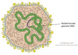
The genome of a fuselloviridae is non-segmented and contains a single molecule of circular, double-stranded DNA. The DNA is positively supercoiled. The complete genome is 15500 nucleotides in length. (source: ICTVdB)
Virion Structure of a Fuselloviridae

Fuselloviridae virions consist of an envelope and a nucleocapsid. The capsid is enveloped. Virions are spindle-shaped, flexible, and have protrusions that extend through the envelope. One pole has short tail-like fibers attached to it. The virions are 100 nm in length and 60 nm in diameter. (sources: ICTVdB, Wiedenheft et al.)
Reproduction Cycle of a Fuselloviridae in a Host Cell

The basic replication cycle of crenarchaeal viruses, including Fuselloviruses, has yet to be determined. There are, however, some trends which have been reported. It is thought that members of the Fuselloviridae family, as well as members of the Rudiviridae, Lipothrixviridae, and Guttaviridae families, associate with host cells by tail fibers which are present on one or both ends of the virion. Some viruses integrate their genome into the host cell's chromosome, while others maintain their genomes as extrachromosomal elements.

Assembly and release of most known crenarchaeal viruses, do not require cell lysis. Most crenarchaeal viruses, twith the exception of the Bicaudaviridae member ATV, appear to set up chronic infections, either continually producing virus particles or doing so in short events resulting in growth inhibition. These long-term chronic infections are thought to be an adaptation to the extremely hot and acidic environments crenarchaeal viruses inhabit.
Viral Ecology & Pathology

As mentioned above, viruses in the Fuselloviridae family infect the archaeon Sulfolobus.

Minggu, 16 Oktober 2011

Virus Famili Coccolithoviridae

Coccolithovirus is a giant double-stranded DNA virus that infects Emiliania huxleyi, a species of coccolithophore[1]. Its genome is 407,339 base pairs long with a G+C content of 41.1%, and contains 472 predicted coding sequences.

Wilson and his team at the Marine Biological Association (MBA), University of East Anglia and Plymouth Marine Laboratory (PML), first observed the virus in 1999. Later in the summer of 2005 researchers at the Plymouth Marine Laboratory (Willie Wilson et al.) and at the Sanger Institute (Holden et al.) sequenced the genome for the EhV-86 strain finding it to have 472 protein-coding genes making it a "giant-virus", and the largest known marine virus by genome.

From initial investigation of the Coccolithoviruses genome, a sequence of genes responsible for production of ceramide was discovered. Ceramide is a controlling factor in cell death, and it is currently thought that Coccolithovirus uses this to prolong the life of Emiliania huxleyi while it uses the host cell to replicate. This is a unique ability unseen in any other viral genome to date.

Notes

    ^ Giant viruses in the oceans: the 4th Algal Virus Workshop Virology Journal 2005
    ^ Sanger institute home for Emiliania huxleyi virus 86
    ^ Giantviruses.org top viruses by genome size.
    ^ Plymouth Marine Laboratory press release.

Virus Famili Baculoviridae

The Baculovirus is from a family of large, rod shaped viruses. It is easily broken down into two major subgroups, according to the nucleocapsoidal arrangements within the actual cell itself. Although the virus does not typically affect humans directly, it can be seen in major forms of scientific research, including artificial protein production as well as vaccinations.

Infection from this virus, most often occurs within insect species feeding of the the leaves of infected plants. Infection typically occurs within young, near larval moths. Fortunately enough, this particular virus is not known to inhabit mammalian lifeforms.

This particular virus is also responsible for the historical 'wilting disease' of the silkworm during the sixteenth century. Seen below is a close up microscopic photo image of the virus.
Baculoviruses menginfeksi larva serangga, menyebabkan infeksi yang akhirnya berubah organisme inang menjadi cair sebagai virus baru yang dilepaskan ke lingkungan. Keluarga virus telah dikenal selama ratusan tahun, dengan rekening awal yang tentang "layu" cacing sutra Cina di abad ke-16. Pada awal abad 20 infeksi virus ditemukan sebagai penyebab dari "layu," dan pada tahun 1947 berbentuk batang virion yang merupakan karakteristik baculoviruses pertama kali terlihat. Ada beberapa diskusi saat ini mengenai apakah genera dalam keluarga Baculoviridae harus direklasifikasi. Alasan untuk hal ini akan dibahas pada bagian berikutnya.

Genom baculovirus adalah non-tersegmentasi dan berisi molekul DNA melingkar, beruntai ganda. Urutan genom lengkap 80000-180000 nukleotida panjang. Diselingi seluruh genom adalah bagian dari sekuens berulang DNA yang dikenal sebagai daerah homolog, atau jam. Struktur kompleks ini dibentuk oleh jam mengulangi 60bp, dengan mengulangi masing-masing berisi sebuah palindrom 28bp-lama tidak sempurna. Daerah homolog ini meningkatkan transkripsi dini serta bertindak sebagai asal-usul untuk replikasi DNA. Banyak dari gen dalam genom baculovirus tumpang tindih di ujungnya, yang memungkinkan sejumlah besar gen yang akan dikodekan dalam jumlah kecil DNA.
Analisis terakhir dari urutan genom baculoviruses menunjukkan bahwa taksonomi baculoviruses perlu diubah. Lebih khusus, telah ditemukan bahwa filogeni dari baculoviruses lebih erat berkaitan dengan klasifikasi organisme inang dari ciri-ciri morfologi virus, yang telah digunakan sebelumnya untuk mengklasifikasikan baculoviruses. (Sumber:. Jehle dkk dan ICTVdB)

Struktur Viron Baculovirus suatu


Virion baculovirus memiliki struktur yang kompleks yang terdiri dari sebuah amplop dan sebuah nukleokapsid berbentuk batang. Kapsid adalah 200-450nm panjang, dan 30-100nm diameter. Kapsid memiliki simetri heliks.


Ketika baculoviruses yang ekstraseluler, mereka dapat ditemukan dalam dua bentuk: virus bertunas (BV) dan virus tersumbat (OV). OVs adalah matriks protein polyhedral atau oval berbentuk kristal di mana satu atau beberapa virion dewasa yang tertanam. Para OVs yang besar, berukuran 0.15-15μm panjang. OV partikel terbentuk di dalam sel yang terinfeksi dan dilepaskan ketika sel lyses. Matriks protein kristal dari OVprotects virus sementara di lingkungan ekstraselular, karena ini, OVs digunakan untuk transfer dari virus antara host.


Dua genera dalam Baculoviridae keluarga definied oleh struktur yang berbeda OV mereka. OVs Granulovirus hanya berisi satu virion, dan tidak memiliki amplop polyhedral (dikenal sebagai kelopak a). Ini OV kecil, memberikan "butiran" penampilan ketika OVs banyak terlihat bersama-sama. Protein yang membentuk matriks kristal OVs Granulovirus dikenal sebagai granulin. OVs Nucleopolyhedrovirus jauh lebih besar daripada OVs Granulovirus, memegang 20 atau lebih virion di setiap partikel OV. Para virion baik terpisah atau digabungkan bersama-sama dalam sebuah kelopak. Protein yang membentuk matriks kristal OVs Nucleopolyhedrovirus disebut polyhedrin.


BVS digunakan untuk sel-sel transmisi dalam inang terinfeksi. BV partikel terdiri dari kapsid tunggal tertutup dalam amplop yang kapsid mendapatkan ketika itu "tunas" keluar melalui dinding sel dan ke dalam sistem host. Tidak seperti OVs, BVS tidak dapat bertahan hidup di luar organisme inang.

OV, yang hadir di lingkungan, ditularkan melalui konsumsi. Sebuah larva serangga mengkonsumsi partikel OV, yang masuk ke dalam midgut larva itu. Ada sifat yang sangat alkali midgut larut matriks protein OV itu, melepaskan virion. Para virion melekat pada sel-sel epitel dan menginfeksi midgut mereka, dengan menggunakan DNA sel inang mekanisme replikasi untuk mereplikasi dirinya sendiri. BV partikel tunas keluar melalui membran sel dan menyebar ke seluruh organisme inang, menyebabkan infeksi sekunder. Para BVS akhirnya menginfeksi organsim seluruh host. Setiap sel yang terinfeksi oleh BV menciptakan sejumlah besar OVs, yang dirilis melalui lisis sel. Karena kenyataan bahwa begitu banyak sel-sel pecah pada saat yang sama, larva dikurangi menjadi cairan putih susu (proses yang dikenal sebagai pencairan). Akhirnya, larva pecah, melepaskan OVs ke lingkungan.


Perlu dicatat bahwa tidak semua baculoviruses yang mematikan. Beberapa baculoviruses mengatur infeksi persisten atau bahkan laten di host sehat. Penularan baculoviruses tersebut adalah vertikal antara host.

Baculoviruses menginfeksi serangga. Sebagian besar infeksi terjadi pada spesies yang terkait erat serangga, seperti Lepidoptera. Baculoviruses mencapai penyebaran secara pasif berinteraksi dengan serangga lainnya. Predator yang mengkonsumsi serangga terinfeksi, karena fakta bahwa lingkungan asam perut mereka tidak akan melarutkan matriks protein sekitarnya OVs, membubarkan virus melalui buang air besar. Tanah merupakan reservoir untuk virus. Jika OVs dapat menghindari radiasi UV matahari dan kondisi basa, maka mereka dapat tetap di dalam tanah untuk waktu yang sangat lama.


Sabtu, 15 Oktober 2011

Famili Adenoviridae

Adenoviruses infect birds, humans, and many other mammals and other organisms. They usually cause respiratory infections, but they can also cause conjunctivitis, gastroenteritis, cystitis, and rash illness. Adenoviruses are divided into four genera: Aviadenovirus (which infects birds), Mastadenovirus (which infects mammals), Atadenovirus and Siadenovirus (which infect a variety of organisms). (sources: CDC, Davison et al.)
Currently, research is being done to see if adenoviruses could be used as a treatment for cancer. After going through extensive engineering to remove the genes which control viral replication (which also creates room to insert genes which are of therapeutic interest), adenoviruses become a promising agent for killing tumor cells. (For more information on the use of adenoviruses in cancer treatment, see Curiel and Dani)
Genome Structure
Adenoviruses have linear, non-segmented, double stranded DNA which is around 30-38kbp (though the size varies from group to group). The complete genome is 35800-36200 nucleotides long. A 55kD virus coded terminal protein is covalently attached 5′ end of each strand. The terminal sequences of each strand are inverted repeats. Because of this, the denatured single strands can form “panhandle” structures. (sources: ICTVdB, Davison et al.)
Virion Structure of a Adenovirus
Adenoviruses are non-enveloped icosahedral viruses measuring 60-90nm in diameter and containing double-stranded DNA. The structure of an adenovirus virion is simple, consisting of a capsid, fibers, a core, and associated proteins. A protein core inside the outer icosahedral protein capsid contains the genome. The capsid is comprised of 252 capsomers (12 of which are pentons, 240 of which are hexons). Trimeric fibers project from the surface and are attached to a penton. (sources: ICTVdB, Davison et al.)
Reproduction Cycle of an Adenovirus in a Host Cell
The life cycle of an adenovirus is divided into early and late phases, separated by the DNA replication process. In the early phase, the virus attaches to a cell with its fibers. The penton base protein interacts with the host cell integrins, and the penton is internalized by the host cell through receptor-mediated endocytosis. The penton is disassembled as it is transported to the nucleus, where the viral particle releases its DNA. The viral DNA takes over as terminal protein attached to the end of the DNA strand initiates transcription. The early genes are responsible for making regulatory proteins, which alter the host proteins to prepare for DNA synthesis, activate other virus genes, and provide protection from the host’s immune system. Viral DNA replication now occurs.
The late phase begins when the late genes are expressed during DNA replication. These genes produce proteins that are involved in virus particle assembly. The host’s cellular processes are shut down as transport of mRNA to the cytoplasm is blocked when the late genes are expressed. Viral mRNA is transported to the cytoplasm and translated instead. Virus assembly occurs. Finally, the host cell dies due to a build-up of adenovirus death protein. As the cell dies, the new virus particles are released from the cell. (sources: Viral Bioinformatics Research Center, ICTVdB)
Viral Ecology & Pathology
Adenoviruses infect many different species in many different ecosystems. According to the Centers for Diesease Control and Prevention, they can survive for long periods of time outside the body due to an unusually high resistance to chemical or physical agents and adverse pH conditions. Transmission is usually through direct contact, fecal-oral transmission, or waterborne transmission.